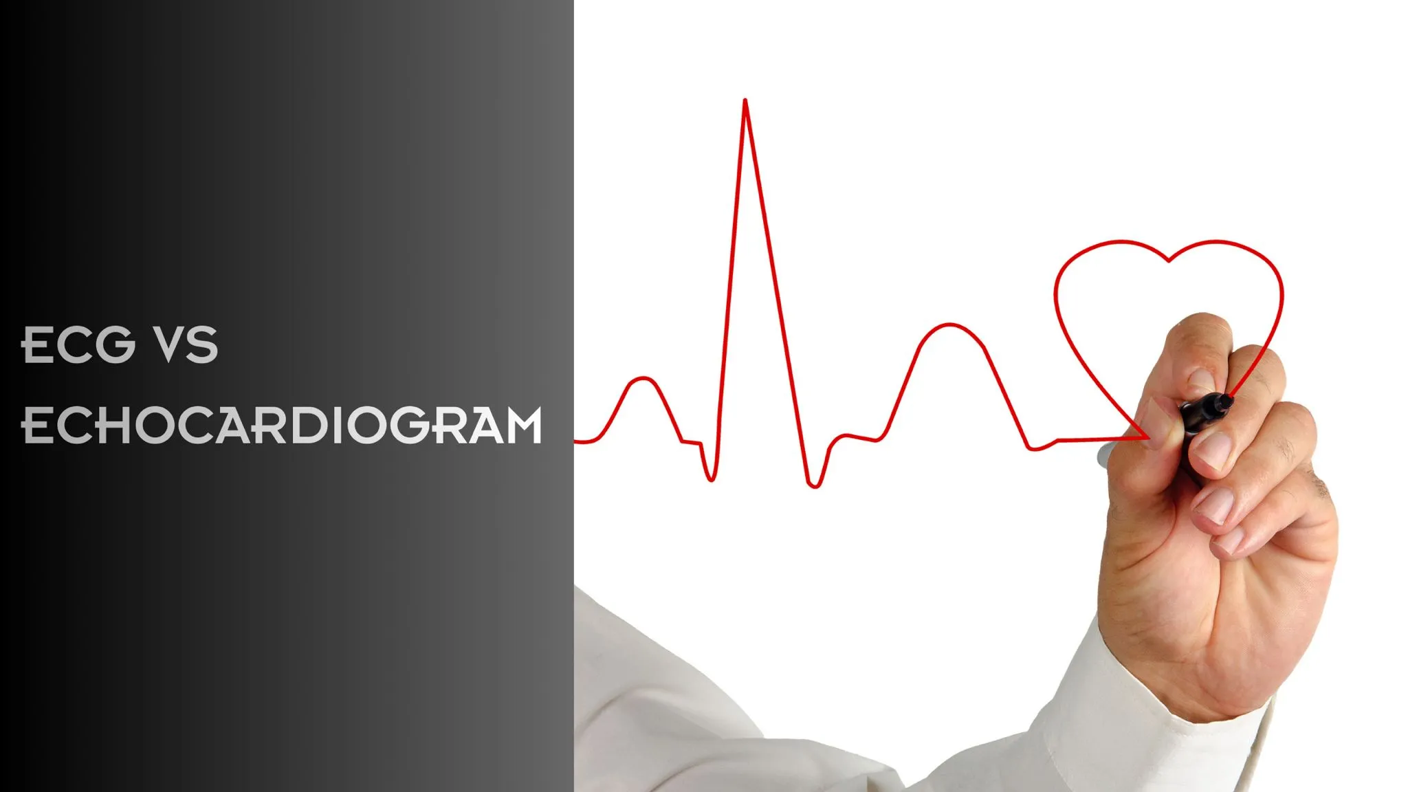
ECG Vs Echocardiogram: Difference Between ECG and Echo
Has your cardiologist suggested an ECG or an Echocardiogram, but you’re unsure whether they are the same or different, or whether you need both tests to understand your heart health? Some people confuse an electrocardiogram with a heart echo and assume they are the same. However, they serve different purposes in evaluating your heart health.
Both an ECG and an echocardiogram are important diagnostic tests used to monitor and assess your cardiac wellbeing. Cardiologists often recommend these tests when someone experiences unusual symptoms, has a family history of heart disease, or is being monitored for a specific cardiovascular condition.
In this guide, we explain ECG vs Echocardiogram – purpose, procedure, and uses of each test to help you understand the key differences between the two.
What is an Electrocardiogram (ECG)?
An electrocardiogram is the most basic yet important heart test that records your heart’s electrical activity and displays it as a graph. Each heartbeat is triggered by electrical signals that control the contraction and relaxation of heart muscles. The ECG captures these signals and displays them as a graph, allowing doctors to evaluate your heart rhythm and determine whether it is normal or irregular.
Book a Private ECG Test in London
How is the ECG Test Performed?
During an electrocardiogram, you’ll be asked to lie straight. Small adhesive electrodes are placed on your chest, arms, and legs, and then connected to the ECG machine. The sensors detect the heart’s electrical activity and record it as an ECG waveform.
What Does an Electrocardiogram Show?
An ECG is usually performed to detect the following heart conditions:
- Heart rhythm disturbance
- Previous heart attack
- Restricted or poor blood flow
- Bradycardia (Slower than normal heart rate ) and Tachycardia (Faster than normal heart rate)
What is an Echocardiogram (Echo)?
An echocardiogram is an ultrasound scan of the heart that produces real-time images to evaluate its structure and function. It is completely different from an ECG, which monitors the heart’s electrical activity. Echocardiography helps cardiologists analyse the heart’s structure, pumping efficiency, blood flow, and overall cardiac performance, thereby enabling the diagnosis of structural and functional heart problems.
How is an Echo Scan Performed?
During an echocardiogram, you’ll lie straight on an examination couch. A small amount of gel is applied to an ultrasound probe to help it move frictionlessly across your chest and capture real-time images of the heart’s structure and performance. The probe transmits sound waves into the chest, and the reflected waves are used to generate images that help cardiologists identify structural and functional defects.
What Does an Echocardiogram Show?
A heart echo can detect various cardiac conditions, such as:
- Cardiomyopathy
- Valvular Heart Disease
- Congenital Heart Defects
- Pericardial Effusion
- Previous Heart Attack
- Heart Failure
ECG Vs Echocardiogram: What are the Key Differences?
Both an ECG and an echocardiogram are essential heart tests, but they measure different aspects of your heart’s health. Understanding how each test works and what it reveals can help you see why your doctor might recommend one or both for a complete assessment of your heart. Here are some key differences explained for your understanding:
| Electrocardiogram (ECG) | Echocardiogram (Echo) |
| Measures heart rate and electrical activity | Provides real-time images of the heart structure and function |
| ECG uses electrodes and electrical recording | An echocardiogram uses ultrasound imaging |
| Helps detect arrhythmias and other rhythm disorders | Helps detect structural defects and perform functional assessment |
| Displays results in the form of a waveform graph | Provides insights with real-time heart images |
| Can not show the heart structure and muscle thickness | Identifies structural abnormalities, valvular disease, and congenital defects |
| Often used in an emergency for quick assessment | Used for detailed evaluation of chronic conditions |
| Helps detect heart rhythm disorders and prior heart attacks | Helps diagnose heart failure and cardiomyopathy |
| An ECG typically takes around 5-10 minutes | An echocardiogram takes around 20-30 minutes |
EKG or Echocardiogram: Which Test Do You Actually Need?
The type of heart test you need depends on your symptoms, medical condition, and what your doctor is trying to assess. An ECG is commonly recommended to identify underlying causes of heart rhythm disturbances, palpitations, chest discomfort, or arrhythmias.
An echocardiogram, on the other hand, is used to detect structural and functional abnormalities of the heart. In many cases, these tests are complementary rather than interchangeable. Your cardiologist may recommend one or both to obtain a comprehensive and accurate understanding of your cardiac health.
Are There Any Risks?
An electrocardiogram and a heart echo are completely safe, non-invasive, and painless tests. No harmful radiation or sedation is involved in either. With an ECG, the electrodes may cause slight temporary discomfort, which typically resolves on its own. You can return home immediately after the procedure and resume your normal activities without any restrictions.
Learn: Why Early Heart Screening Is Crucial?
When Can You Expect Results?
ECG results are usually available the same day, often within a few hours. Echocardiogram results may take slightly longer, depending on report review time and your doctor’s availability to discuss the findings.
Bottom Line
Hopefully, this quick guide will help you understand the difference between an ECG and an echocardiography. Both tests are safe, painless, and essential for diagnosing a wide range of heart conditions. If you’re in London or nearby and want expert evaluation, you can book an appointment with Francesco Lo Monaco, a specialist in advanced heart imaging and diagnostics, for a personalised assessment and care.
FAQs
Why is an ECG done before an echo?
An electrocardiogram (ECG) is usually performed first because it is a simple, quick, and important diagnostic test that helps identify immediate heart rhythm or electrical disturbances. It provides a rapid initial assessment, helping physicians determine whether further evaluation is necessary.
Is an echocardiogram necessary if the ECG is normal?
If your ECG report is normal, there is usually no need for an echocardiogram. However, an echocardiogram may still be recommended if you are experiencing symptoms such as unexplained fatigue, shortness of breath, or chest discomfort, or if your physician suspects an underlying cardiac condition that an ECG cannot detect.
What does an echocardiogram show that an EKG does not?
An ECG (EKG) only shows the heart’s electrical activity and rhythm. An echocardiogram provides real-time images of the heart’s structure, including its chambers, valves, muscle movement, and pumping function, which an ECG cannot show.
Which is more reliable, an echocardiogram or an ECG?
For detailed structural and functional insights into your heart health, an echocardiogram is considered more accurate and reliable. An ECG is typically performed during the initial assessment to evaluate the heart’s electrical activity and rhythm.
Is an ECG or an echo scan more costly?
An echocardiogram is a more advanced, costly cardiac test that evaluates the heart’s structure, function, and pumping efficiency in real time.