PRIVATE ECHOCARDIOGRAM IN LONDON- HEART SCAN
£390
Rapid Access to Cardiology Tests and Scans
State of the Art Diagnostics
With Results Available within 48 hours
Wide-Ranging Cardiac Expertise
WHAT IS ECHOCARDIOGRAPHY?
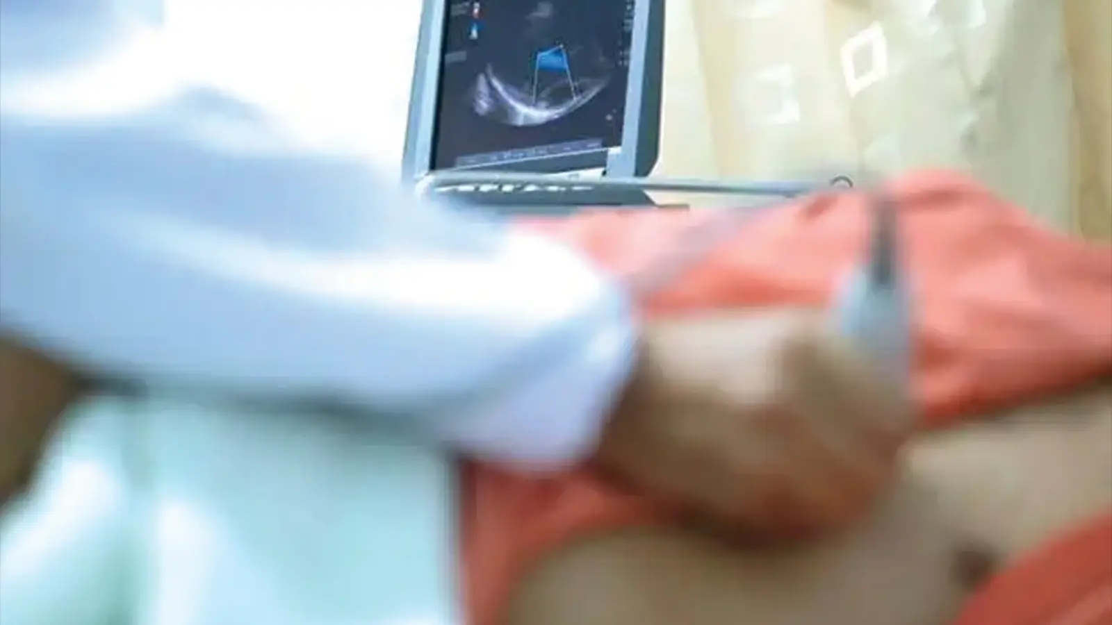
A transthoracic echocardiogram or heart echo is a safe, painless, and non-invasive ultrasound that uses sound waves to create real-time images of your heart to analyse the heart’s structure, rhythm, and blood flow and detect any abnormalities.
WHAT HAPPENS DURING AN ECHO HEART SCAN?
During a heart echo, the technician or cardiologist places a small handheld transducer on your chest to record detailed ultrasound images of your heart. This device visualises the chambers, valves, and overall heart function in real time. The procedure takes around 20–30 minutes and provides accurate insights into any structural or functional abnormalities.
TYPES OF ECHOCARDIOGRAM HEART SCAN
Stress Echo
A Stress Echocardiogram evaluates how your heart functions under physical stress. It is especially useful for detecting coronary artery disease or restricted blood flow that may not be visible at rest.
Transoesophageal Echocardiogram (TOE)
A TOE scan provides clear visuals of heart valves and chambers using a probe that is passed gently down the throat.
Foetal Echocardiogram
This test examines the fetal heart to identify structural or rhythm abnormalities, ensuring timely monitoring and treatment if needed.
Bubble Echo
A Bubble Echocardiogram uses imaging ultrasound and sterile saline with microbubble contrast to precisely detect a hole in the heart or any blood flow abnormalities.
Contrast Echocardiogram
This heart echo scan involves injecting a contrast agent into the bloodstream to enhance echocardiographic resolution and detect subtle abnormalities more accurately.
WHY IS A PRIVATE ECHOCARDIOGRAM NEEDED?
A private heart echo test helps doctors detect structural and functional abnormalities in the heart. Early diagnosis and timely treatment can prevent disease progression and further complications, such as:
What an Echo Doesn’t Replace?
A transthoracic echocardiogram does not directly detect blocked arteries or intermittent heart rhythm problems. For long-term heart rhythm monitoring, a Holter Monitor can be the preferred choice. Please remember, an echo scan doesn’t replace an urgent emergency for active chest pain.
How Much Does an Echocardiogram Cost?
Our standard transthoracic echocardiogram costs £390, including the scan and a same-day written report. Self-pay and all major UK insurers accepted.
Pay With Convenience
Self-Pay Patients
Patients who prefer to pay directly can book appointments without insurance. We offer various self-pay options for initial & follow-up consultations and diagnostic tests, including pay-as-you-go, debit or credit card payments, and PayPal.
Insured Patients
If you have private medical insurance, consultations and tests can be covered depending on your policy. We work with most major UK insurance providers, including AXA, Aviva, Bupa, and Vitality.
Book Your Private Echocardiogram in London
Get fast access to specialist-led cardiac ultrasound with Dr Francesco Lo Monaco in London. Same-day appointments, no GP referral required.