PRIVATE EXERCISE TOLERANCE TEST (ETT) LONDON
£350
(Exercise ECG | Stress Test | Treadmill ECG )
WHAT IS AN EXERCISE TOLERANCE TEST (ETT)?
An Exercise Stress Test is a non-invasive cardiac screening that measures how well your heart responds while performing some activity. Monitoring heart rate, rhythm, blood pressure, and ECG patterns helps your cardiologist detect various heart conditions, such as CAD and arrhythmias, as well as evaluate the effectiveness of ongoing treatment.
ALL ABOUT AN EXERCISE TOLERANCE TEST
What Happens During AN ETT Test?
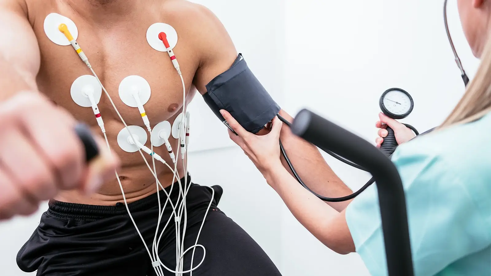
During an Exercise ECG, small adhesive electrodes are attached to your chest and connected with an ECG monitor to analyse your heart’s electrical activity, blood pressure, and any signs of discomfort. The ETT heart test typically lasts for 20 to 30 minutes and is carried out by a male or female cardiac physiologist. You are asked to perform various physical activities, such as walking on a treadmill and cycling, during which the speed and gradient gradually increase. Your vitals are recorded every 3 minutes to assess how your heart responds to this exertion.
Results and Follow-Up Care
Once you stop exercising, your heart rate and blood pressure return to normal. Your cardiologist will review your stress test results based on your vitals, symptoms, and overall response during the procedure. Dr Francesco will then create a personalised treatment plan according to the findings or recommend further tests or scans for more comprehensive insights.
TYPES OF ELECTROCARDIOGRAM (EKG/ECG)
Your heart doctor will recommend the most suitable type of electrocardiogram based on your symptoms and any suspected heart condition.
Exercise Stress ECG
It is the most common and fundamental heart assessment involving physical exertion, while your heart’s electrical activity, BP, and symptoms are monitored through an ECG.
Stress Echocardiogram
Stress Echo combines a standard stress test with comprehensive ultrasound imaging to assess the heart’s structure, pumping efficiency, and blood flow during peak physical activity.
Nuclear Stress Test
The Nuclear Stress Test uses a safe radioactive substance and cardiac imaging to evaluate your heart muscles for identifying the areas with poor blood circulation and arterial blockage.
Cardiopulmonary Exercise Test
The CPET measures how your heart, lungs, and muscles respond to physical exertion to diagnose unexplained breathlessness and assess cardiac fitness.
WHY IS AN EXERCISE ECG IMPORTANT TO ASSESS YOUR HEART?
An Exercise Stress ECG Test provides vital assessments for diagnosing, monitoring, and managing various heart conditions while in physical stress, such as:
FAQs
BOOK A CARDIOLOGY APPOINTMENT ONLINE
If you’ve recently had a specialist heart checkup and feel unsure about the diagnosis or treatment plan, getting a second opinion can provide the clarity you need. Schedule a consultation with Dr. Lo Monaco, an expert private cardiologist in London, or book your heart test/scan today for fast, specialist cardiac care.