What Does Atrial Fibrillation Look Like on an ECG?
Atrial Fibrillation is the most common form of arrhythmia (heart rhythm disorder), caused by chaotic, disorganised electrical activity in the atria, the upper chambers of the heart. According to the NHS, around 900,000 people in England are living with Atrial Fibrillation, accounting for approximately 1.74% of the population.
Although common, AFib can be a serious condition that may require urgent medical attention. If left untreated, it can significantly increase the risk of stroke, heart failure, and cardiomyopathy.
Understanding what does atrial fibrillation looks like on an ECG is one of the most important steps towards early diagnosis and protecting your long-term heart health.

Don’t Wait Weeks for Answers
Get fast, specialist-led heart checks with same-day or quick appointments
What is Atrial Fibrillation (AFib)?
Atrial Fibrillation (AFib) is a common heart rhythm disorder, caused by chaotic, disorganised electrical activity in the atria, the upper chambers of the heart. It occurs due to a glitch in the heart’s electrical system that triggers multiple abnormal electrical signals within the atria. As a result, instead of contracting in a coordinated sequence, the atria quiver rapidly, sending erratic electrical signals to the ventricles below.
This disrupts the heart’s normal pumping efficiency and, if left untreated, significantly raises the risk of:
- Stroke
- Heart Failure
- Cardiomyopathy
- Blood Clot Formation
Common Symptoms of AFib
Atrial Fibrillation doesn’t always cause noticeable symptoms – in fact, some patients only discover the condition during a routine ECG. However, when symptoms do occur, they typically include:
- Heart palpitations
- Weakness and reduced exercise tolerance
- Dizziness or fainting
- Shortness of breath
- Chest pain
If you are experiencing any of these symptoms, an ECG is an essential first step in determining whether AFib is the underlying cause.
How Does an ECG Record Heart Rhythm?
An electrocardiogram (ECG) is a non-invasive test that records the electrical activity of the heart through electrodes placed on the skin. Each heartbeat generates a distinct electrical signal, which is captured as a waveform on the ECG graph.
What Does a Normal ECG Look Like?
In a healthy individual with a normal sinus rhythm, the ECG displays a regular, well-organised waveform made of 3 key components:
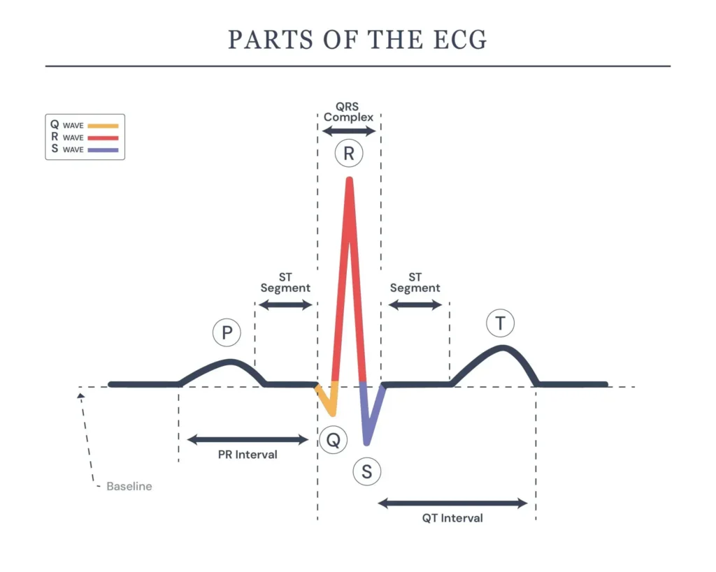
| ECG Component | What it Represents | Normal Appearance |
| P Wave | Atrial Depolarisation | Distinct, rounded peaks before each QRS |
| QRS Complex | Ventricular Depolarisation | Sharp, narrow spike that is less than 120 ms wide |
| T Wave | Ventricular Repolarisation | Broad, rounded wave after each QRS. |
Also Read: What does an abnormal ECG mean?
Atrial Fibrillation ECG Review
Your heart produces an electrical signal that causes your heart to contract and relax for pumping blood throughout the body. This electrical signal is produced by the ‘sinus node’, the heart’s natural pacemaker, which creates a normal heartbeat. The electrical signal travels from the atria to the ventricles in a controlled, well-coordinated pattern.
In AFib, a glitch in the heart’s normal electrical activity causes the atria to beat very fast, causing the ventricles to beat out of sync. These abnormal rhythms can increase the heart rate from a normal range of 60-100 beats/minute to around 100-175 beats/minute. Some people may experience a slower or faster heart rate depending on their condition and treatment.
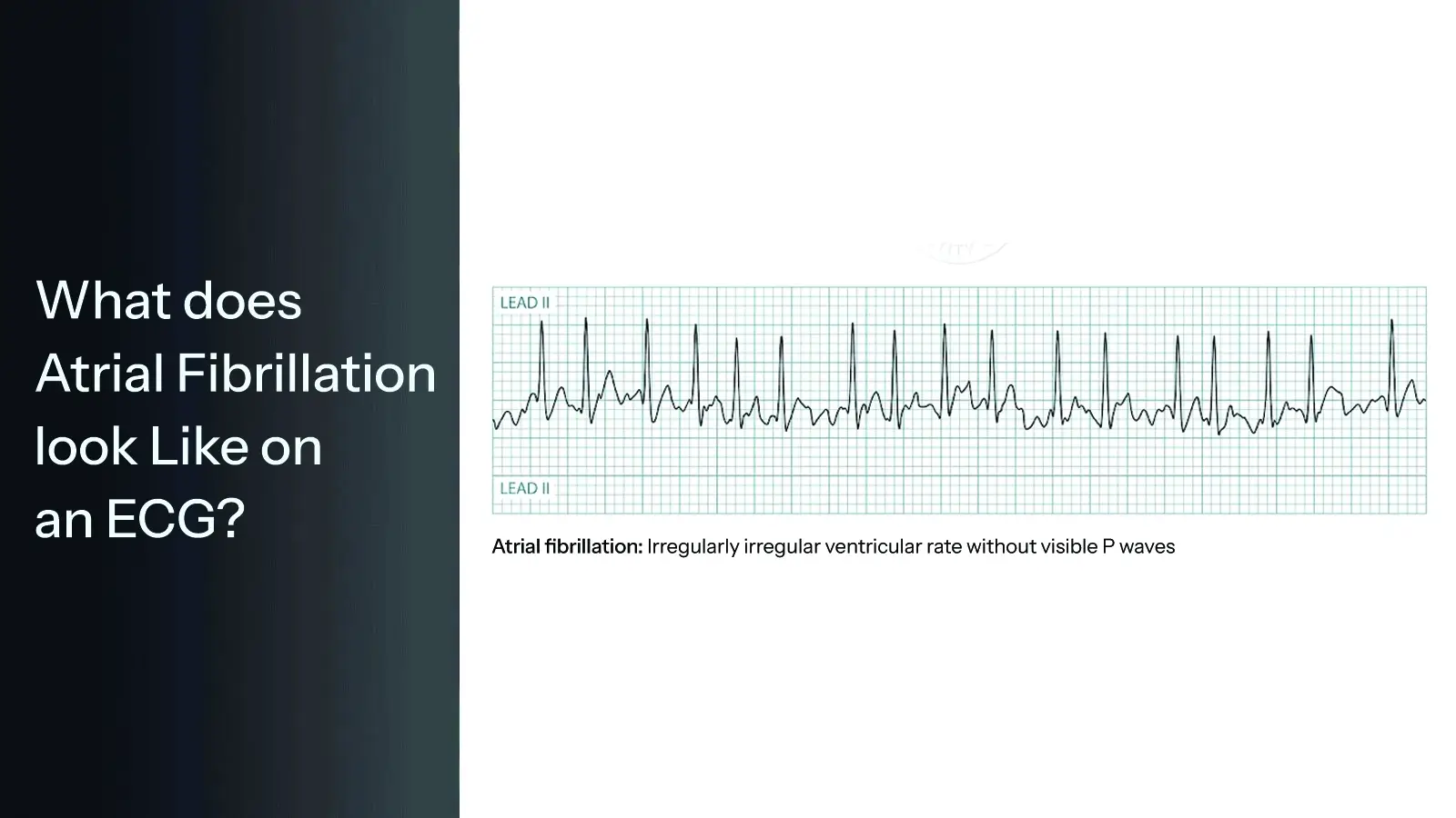
What Does Atrial Fibrillation Look Like on an ECG?
AFib produces an abnormal ECG pattern that a cardiologist can identify at a glance. The key differences from a normal sinus rhythm are:
Absence of Distinct P Waves
In Atrial Fibrillation, the organised electrical signals from the sinus node are replaced by chaotic, rapid impulses firing simultaneously across the atria. As a result, the clearly defined P waves disappear from the ECG. Instead, the baseline between heartbeats appears irregular and uneven.
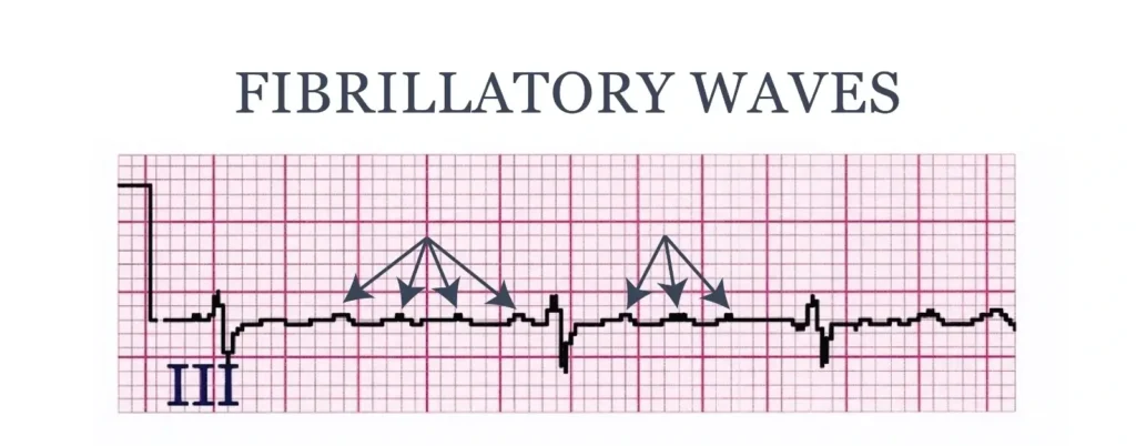
Fibrillatory Waves
Rather than clear P waves, the AFib ECG shows small, irregular waves known as fibrillatory waves. These appear as a chaotic baseline and represent the disorganised electrical activity occurring within the atria.
Narrow QRS Complexes
The QRS complexes (the spikes that represent ventricular contraction) usually appear narrow, but occur at irregular intervals because the ventricles are receiving unpredictable electrical signals from the atria.
Variable Ventricular Rate
The ventricular rate in AFib is generally rapid, ranging from 90 to 175 beats per minute. However, in patients on heart rate-controlling medications, the ventricular rate may appear controlled or even slow.
No Isoelectric Baseline
In a normal sinus rhythm, a flat baseline is clearly visible between waveforms. In AFib, this flat baseline is replaced by the irregular fibrillatory activity.
Normal Sinus Rhythm Vs Atrial Fibrillation
| ECG Feature | Normal Sinus Rhythm | Atrial Fibrillation |
| Heart rhythm | Regular | Irregularly irregular |
| P Wave | Distinct, present before each QRS | Absent, replaced by fibrillatory waves |
| QRS Complex | Narrow | Usually narrow, may be wide when conduction occurs through an aberrant pathway. |
| Baseline | Flat isoelectric line | Chaotic baseline |
| QRS intervals | Consistent | Completely variable |
| Heart rate | 60-100 bpm | Often 90-175 bpm |
Atrial Fibrillation Vs Atrial Flutter
Atrial Fibrillation is a chaotic heart rhythm that originates from the top left chamber of the heart – the atria. Atrial Flutter, on the other hand, is a fast but regular rhythm that originates from the heart’s top right chamber. If left untreated for long, both these heart conditions can cause stroke.
When Should You Have an ECG for Atrial Fibrillation?
We recommend booking an ECG if you experience any of the following:
- Frequent and severe heart palpitations
- Unexplained fatigue
- Breathlessness, particularly during mild activity
- Chest pain/tightness
- Dizziness/fainting
Book a Private ECG and Heart Consultation in London
If you are concerned about your heart rhythm irregularities, you can book a private ECG and consultation with Dr Francesco Lo Monaco, a leading cardiologist in London. Appointments are available on a same-day and next-day basis, with access to advanced facilities, ensuring prompt and accurate evaluation of your heart health.
Disclaimer
The information provided in this blog is for general information and educational purposes only. The content is not intended to be a substitute for professional medical advice, diagnosis, or treatment. Always seek the advice of your GP or a qualified medical professional regarding any medical condition. If you are experiencing a medical emergency, please contact emergency services immediately.